WebPathology(@WebPathology) 's Twitter Profileg
WebPathology

@WebPathology

Visual survey of surgical pathology with more than 13,000 high-quality images of benign and malignant neoplasms & related entities. Created by: @DharamRamnani

ID:171921202

calendar_today28-07-2010 14:01:32

1,6K Tweets

13,9K Followers

1,2K Following

Follow People
WebPathology(@WebPathology) 's Twitter Profile Photo

Testis#3. Young male. Enlarging testis. US→heterogenous, vascular mass. beta-hCG > 30,000 mIU/ml (ref. range 0-3). AFP & LDH within ref. range. Classic macro- and microscopic appearance.

Testis#3. Young male. Enlarging testis. US→heterogenous, vascular mass. beta-hCG > 30,000 mIU/ml (ref. range 0-3). AFP & LDH within ref. range. Classic macro- and microscopic appearance. #GUPath #PathTwitter
account_circle
WebPathology(@WebPathology) 's Twitter Profile Photo

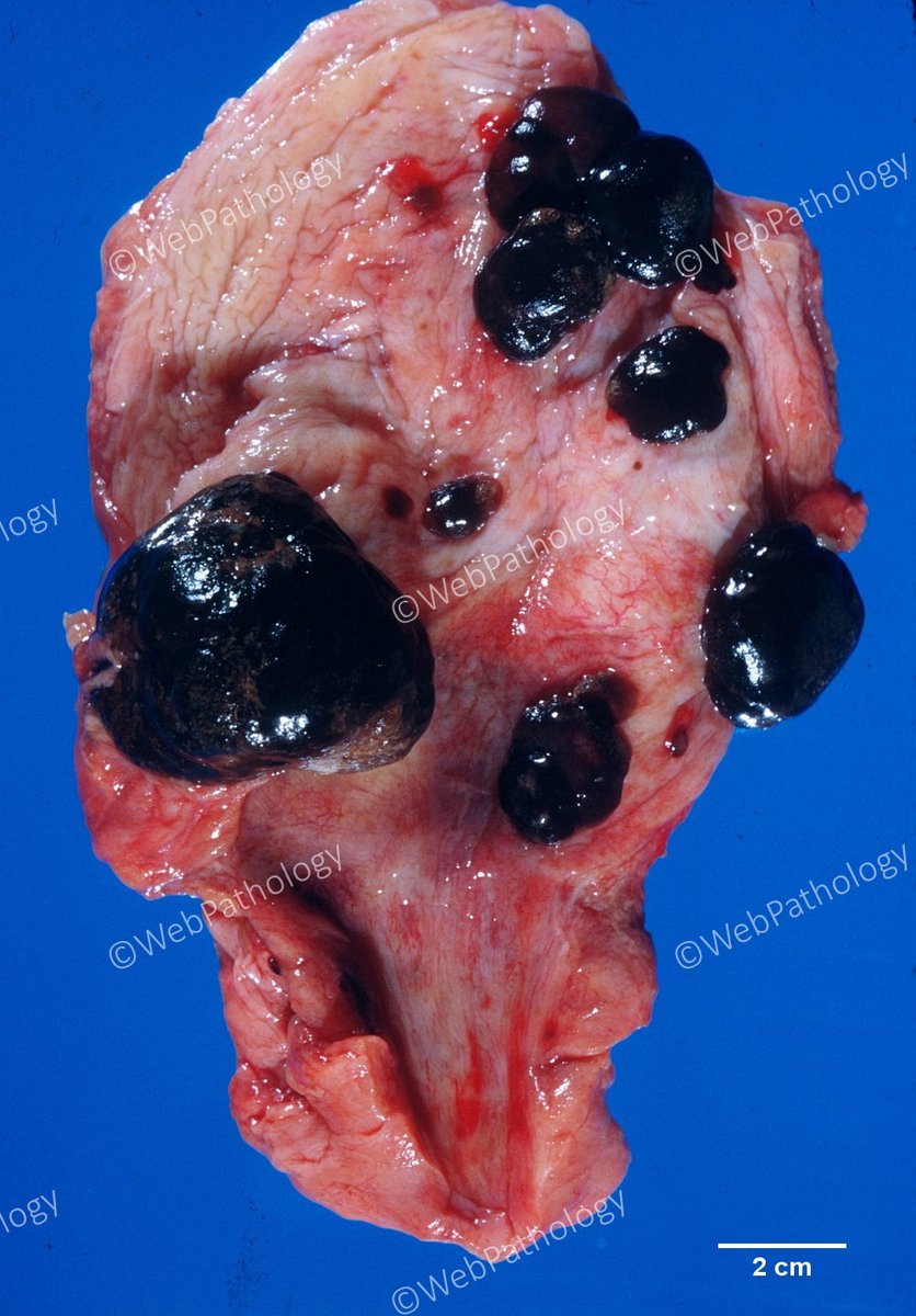
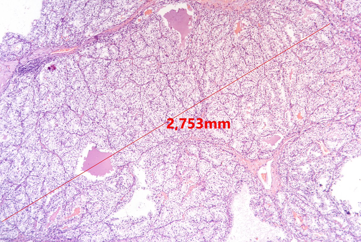
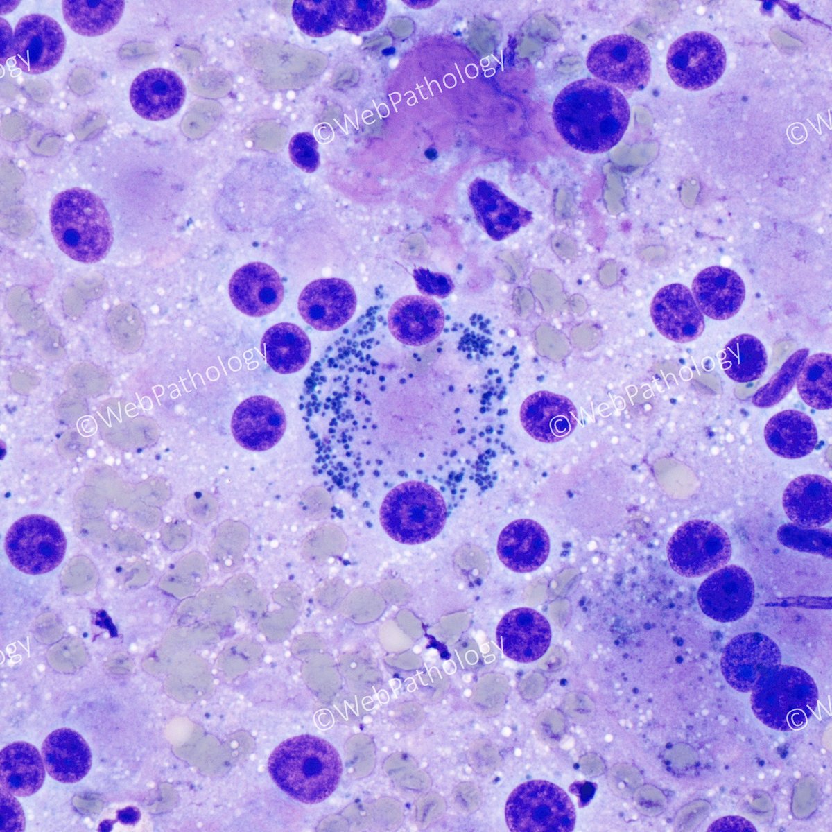
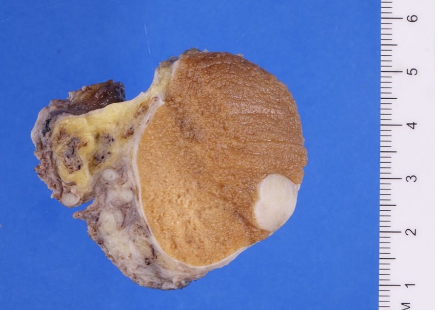
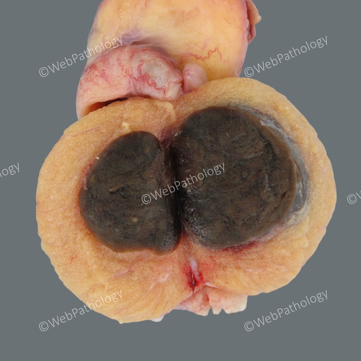
Adult male with infertility, azoospermia, elevated testosterone & testicular mass. Tumor markers (AFP, LDH, hCG) within ref. range. Diagnosis: Leydig Cell Tumor. Dark color due to lipofuscin (seen in 15% cases). Last image - Diff-Quik stained touchprep.

Adult male with infertility, azoospermia, elevated testosterone & testicular mass. Tumor markers (AFP, LDH, hCG) within ref. range. Diagnosis: Leydig Cell Tumor. Dark color due to lipofuscin (seen in 15% cases). Last image - Diff-Quik stained touchprep. #GUPath #PathTwitter
account_circle
WebPathology(@WebPathology) 's Twitter Profile Photo

Testicular Neoplasms. Case 2: Adult male (> 35 yrs) with a testicular mass. Any guesses based on macroscopic appearance? Clinical info. and labs. to follow soon.

Testicular Neoplasms. Case 2: Adult male (> 35 yrs) with a testicular mass. Any guesses based on macroscopic appearance? Clinical info. and labs. to follow soon. #PathTwitter #PathResidents #GUPath
account_circle
WebPathology(@WebPathology) 's Twitter Profile Photo

Mixed Germ Cell Tumor of Testis with predominantly teratoma (80%) and embryonal CA (20%), microscopic foci of yolk sac tumor and syncytiotrophoblastic cells.

Mixed Germ Cell Tumor of Testis with predominantly teratoma (80%) and embryonal CA (20%), microscopic foci of yolk sac tumor and syncytiotrophoblastic cells. #gupath #PathTwitter
account_circle
WebPathology(@WebPathology) 's Twitter Profile Photo

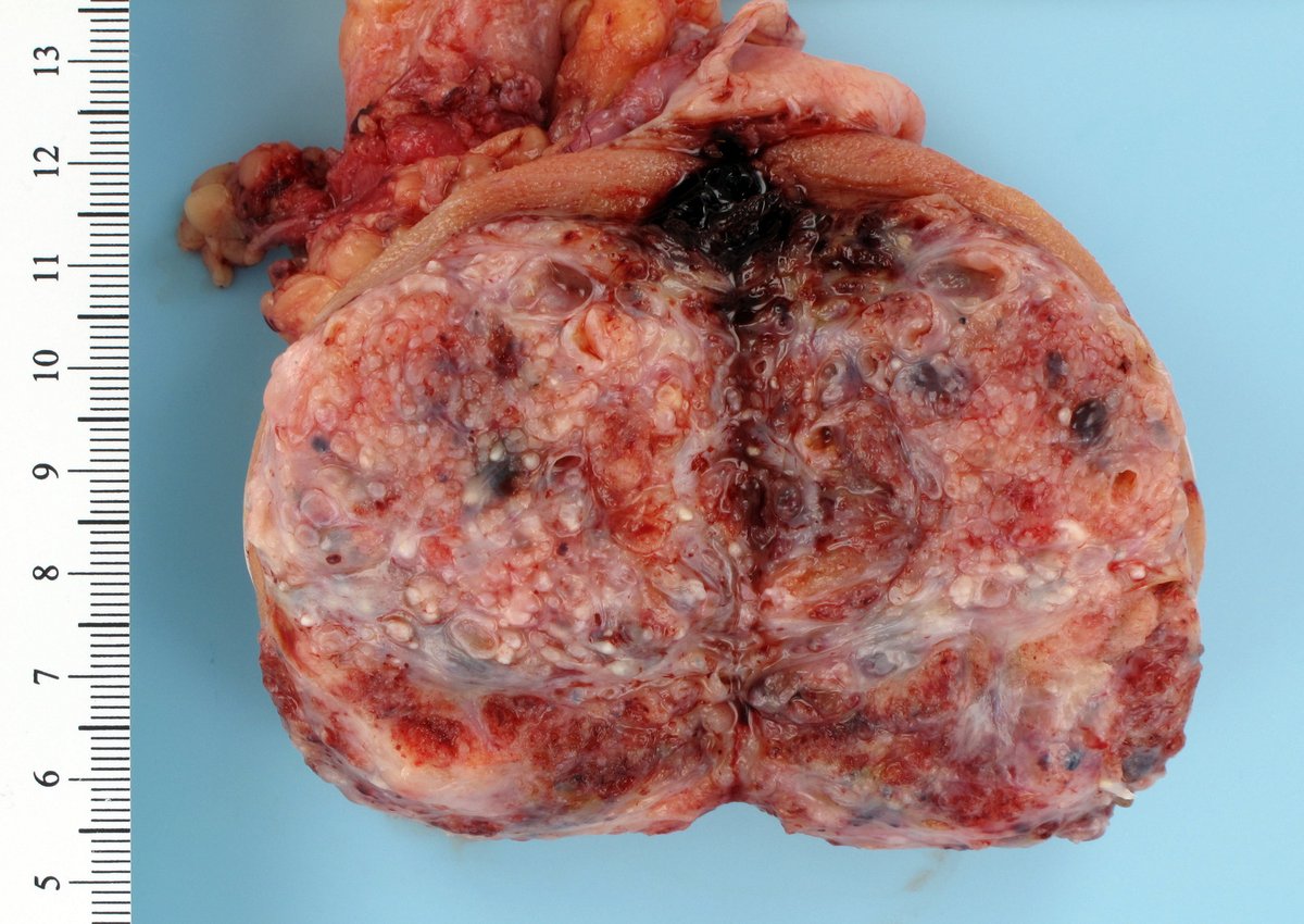
Testicular Cancers. Case 1: Young male, slowly enlarging testis. Ultrasound - complex vascular mass. AFP > 400 ng/ml (0-5.7), hCG > 200 mIU/ml (0-3), LDH > 250 IU/L (121-224). Gross appearance & labs. are classic. Diagnosis & microscopic images tomorrow.

Testicular Cancers. Case 1: Young male, slowly enlarging testis. Ultrasound - complex vascular mass. AFP > 400 ng/ml (0-5.7), hCG > 200 mIU/ml (0-3), LDH > 250 IU/L (121-224). Gross appearance & labs. are classic. Diagnosis & microscopic images tomorrow. #GUPath #PathTwitter
account_circle
WebPathology(@WebPathology) 's Twitter Profile Photo

We see a large number of testicular cancers at my institution. I hope to share some of the interesting cases here over the next few weeks.

account_circle
Katrina Collins, MD(@katcollmd) 's Twitter Profile Photo

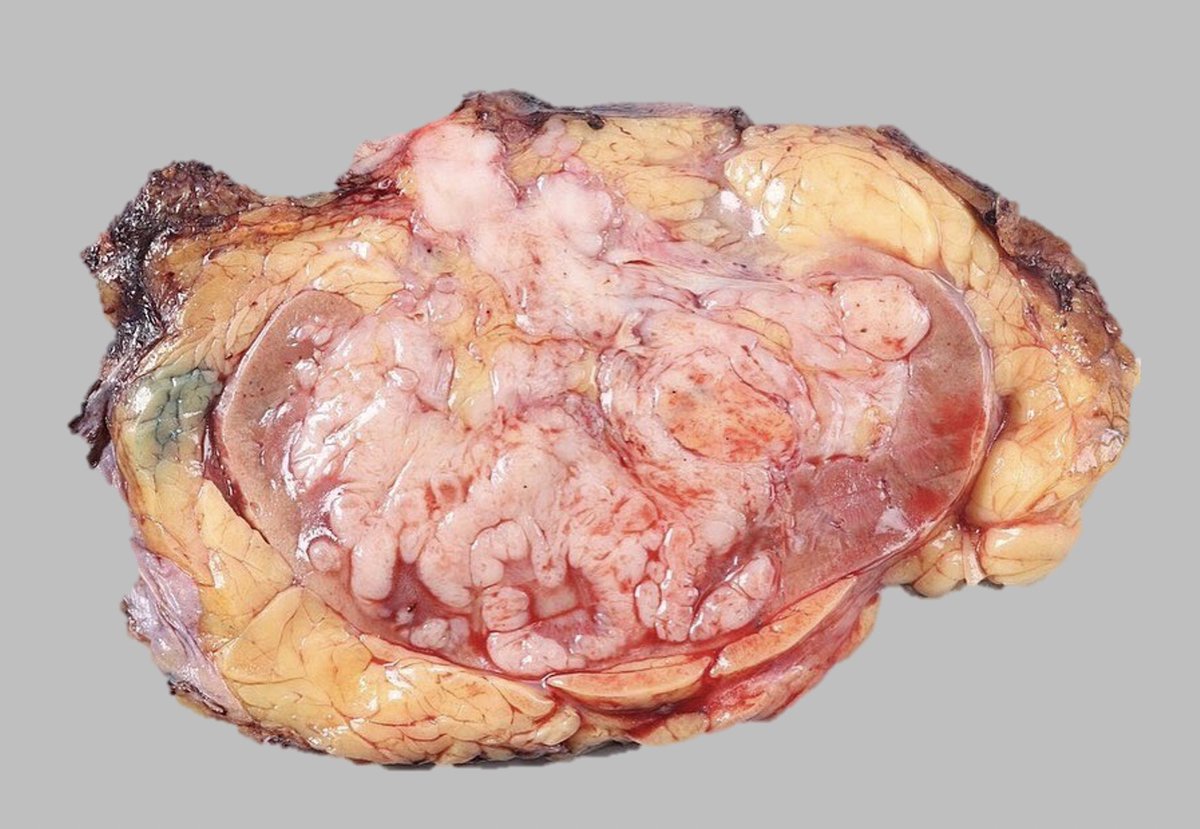
radical nephrectomy, sarcomatoid components comprise sizeable areas displaying dense, tan-pink, or white areas within the tumor, typically with a firm and fleshy texture on cut surface

#macrovsmicro radical nephrectomy, sarcomatoid components comprise sizeable areas displaying dense, tan-pink, or white areas within the tumor, typically with a firm and fleshy texture on cut surface #gupath #pathtwitter #kidneytumor
account_circle
@isaalvaradoc(@vintage81106614) 's Twitter Profile Photo

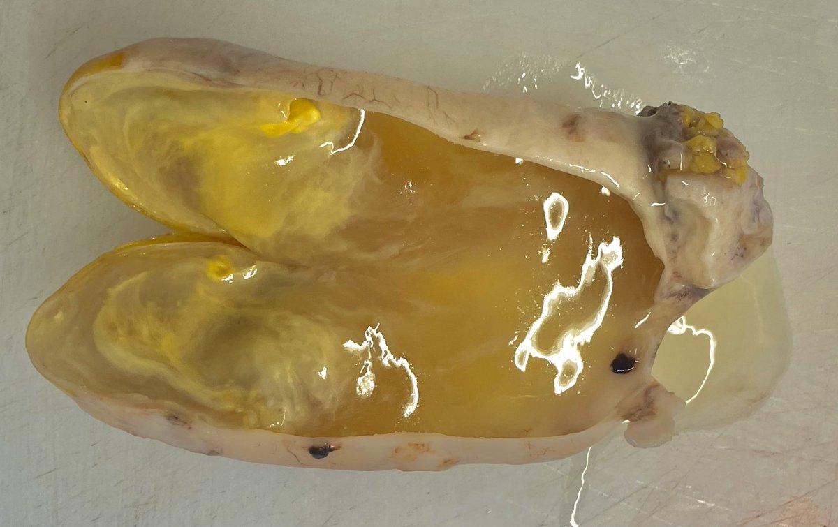
The art of grossing. A 66 yo man with accute- appendicitis like symptoms. Appendix, size:4x2cm, disease restricted to the organ. Final Dx. Low-grade appendiceal mucinous neoplasm (LAMN)

The art of grossing. A 66 yo man with accute- appendicitis like symptoms. Appendix, size:4x2cm, disease restricted to the organ. Final Dx. Low-grade appendiceal mucinous neoplasm (LAMN) #pathology #pathresidents #pathTwitter #GIpath
account_circle
WebPathology(@WebPathology) 's Twitter Profile Photo

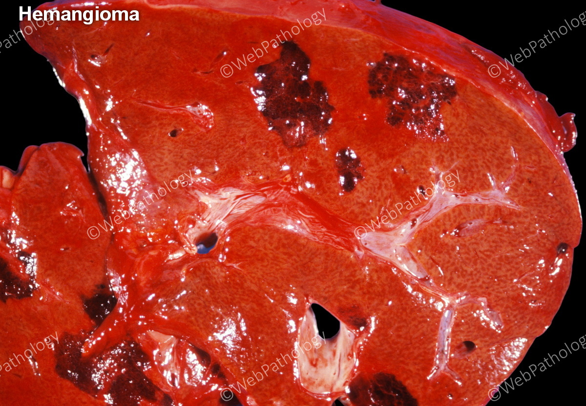
Update in Two of the commonest lesions - Hemangioma & Focal Nodular Hyperplasia have been updated. 50 Images. webpathology.com/case.asp?case=…; up next Hepatocellular Adenoma.

Update in #GIPath #LiverPath Two of the commonest lesions - Hemangioma & Focal Nodular Hyperplasia have been updated. 50 Images. webpathology.com/case.asp?case=…; up next Hepatocellular Adenoma.
account_circle
Steve Vanko, MS, PA(ASCP)(@smvanko) 's Twitter Profile Photo

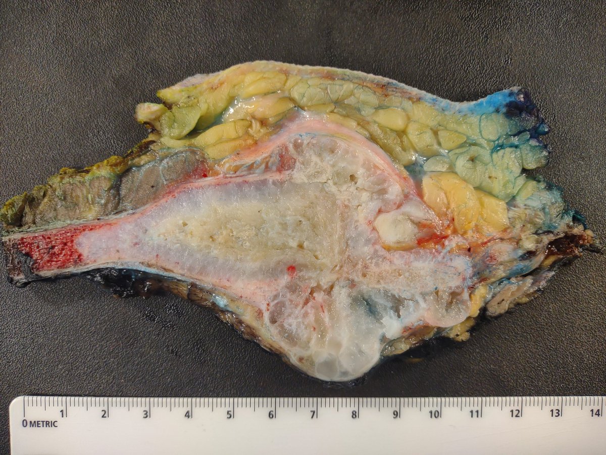
Primary chondrosarcoma of the sternum. Resection includes overlying skin and subcutis , near total sternum with mass and medial aspects of ribs 2-5, pericardium at deep.

Primary chondrosarcoma of the sternum. Resection includes overlying skin and subcutis , near total sternum with mass and medial aspects of ribs 2-5, pericardium at deep. #BST #PathTwitter #GrossPathonX
account_circle