![[n]Beyin(@nBeyin) 's Twitter Profile Photo [n]Beyin(@nBeyin) 's Twitter Profile Photo](https://pbs.twimg.com/profile_images/611172159533264896/BD12qPIm_200x200.jpg)
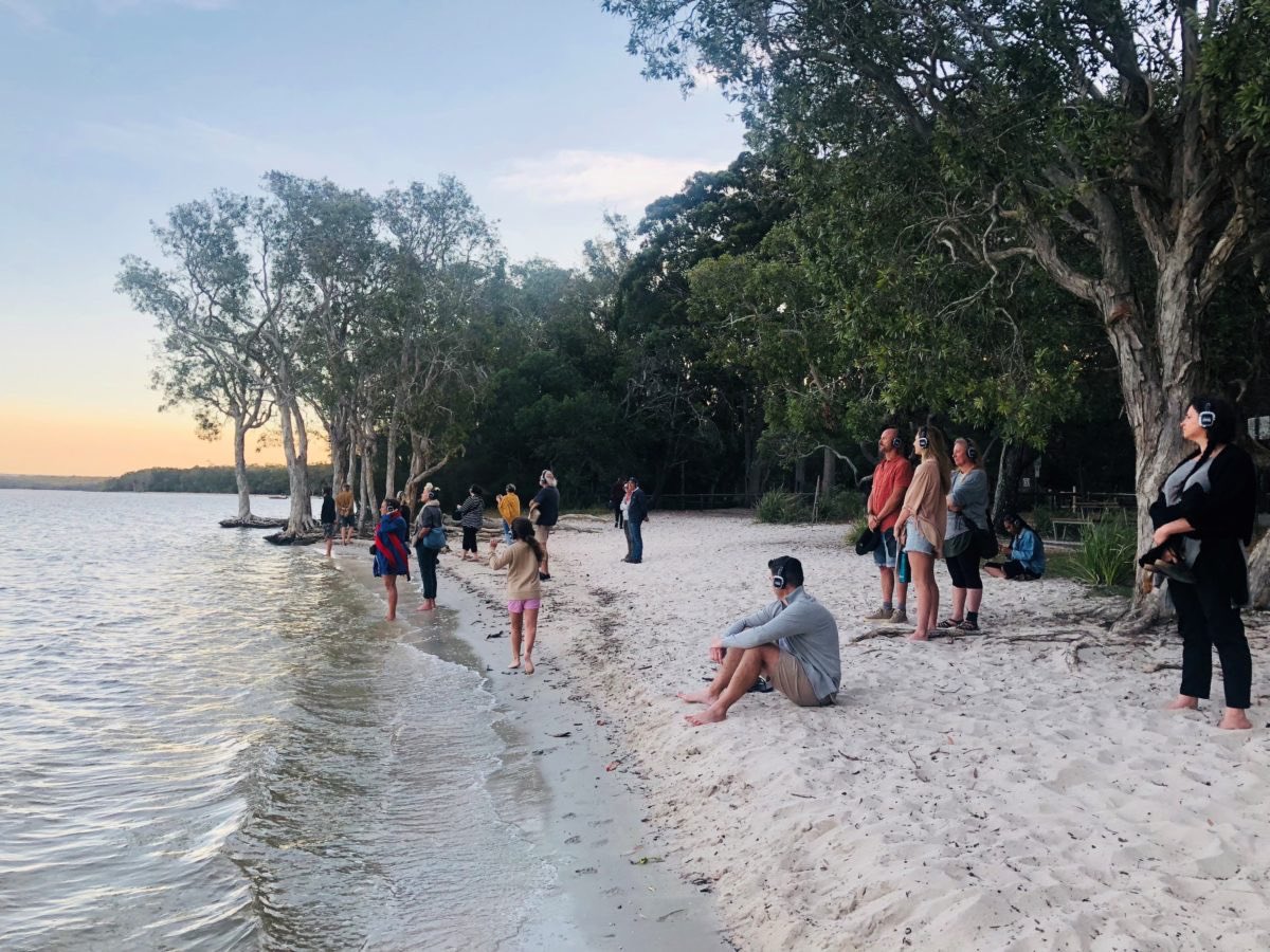
TURN UP THE VOLUME! You are about to explore the world of Biosphere Reserve sounds with Leah Barclay from the BiosphereSoundscapes Project.
Full version available on instagram.com/tv/CBQR8T8FKyl/
#biodiversity #naturelovers

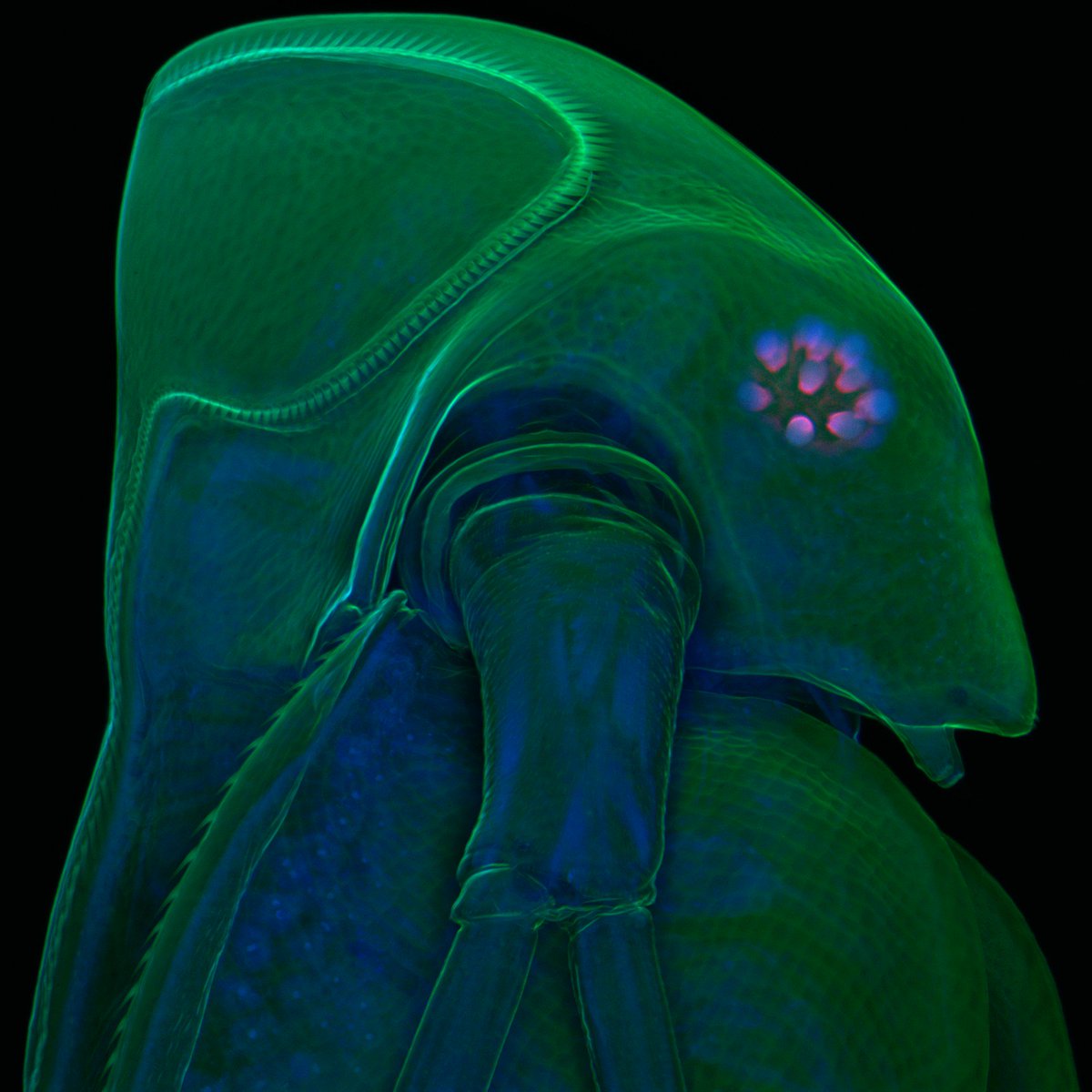
#MicroscopyMonday You can see the exoskeleton of this water flea (Daphnia atkinsoni) in green. But do you see the tiny blue dots? Those are the nuclei of its cells.
🔬📷: Jan Michels, 2009 Olympus BioScapes Digital Imaging Competition
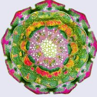
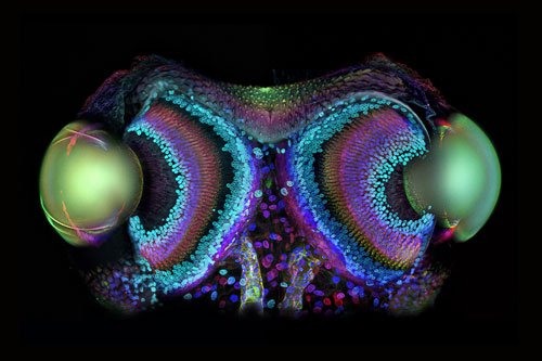
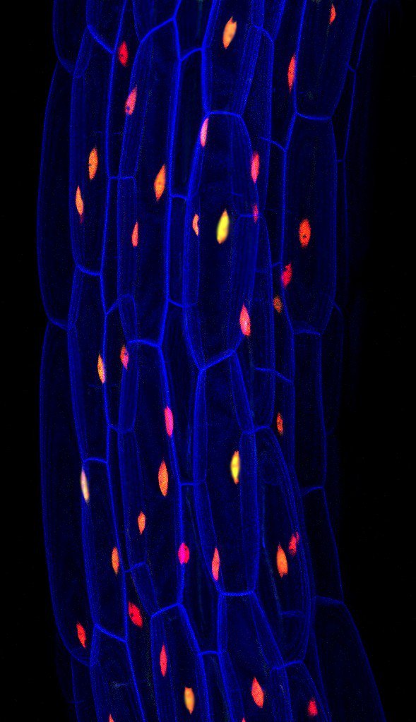
Thale cress seedling!
🔬Confocal
📷Fernán Federici
©Olympus BioScapes
#science #microscopy #microscope #CellBiology #biology

Incredibly exciting new funded PhD at Rutgers, in 'Sound Environments'! sasn.rutgers.edu/academics-admi… Sounding Out! BiosphereSoundscapes hearing places Soundscapes in the Early Modern World Cheryl Tipp amer kanngieser @amkanngieser.bsky.social Northwestern Sound Dr. Jill Rogers Annie Goh Sound Studies Sound@SFU Naomi Waltham-Smith Bill McKibben
Check out Field Trip Symposium tomorrow #environment #sound #fieldtrip #doitfromyourloungeroom BiosphereSoundscapes Horizon Festival artsfront.com/event/42651-fi…

This is a little bit awesome! #soundscape #bioacoustics #mars #soundart #acoustic Acoustical Society of America Sounds Of Urban London BiosphereSoundscapes
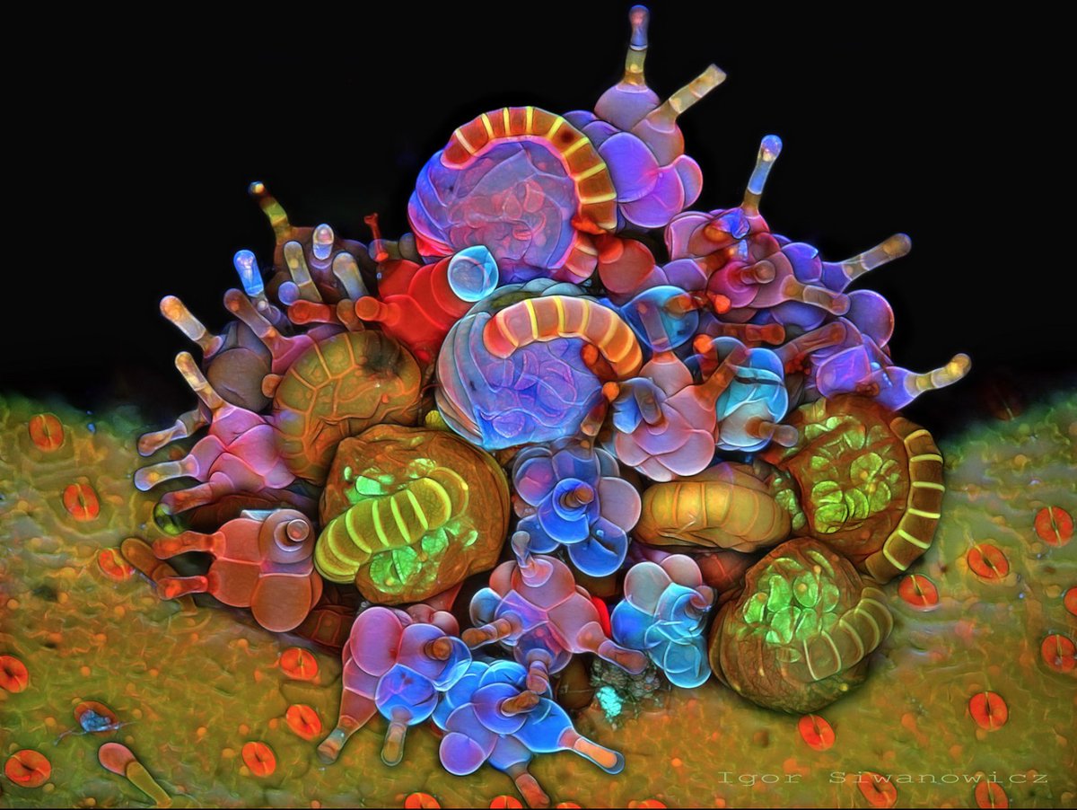
Here are some of Igor Siwanowocz' incredible #microscopy pictures! He is does research at HHMI | Janelia, and is my microscopy idol. He also won many awards at the Nikon Small World
and Olympus bioscapes competitions.
photo.net/gallery/929115…
#BioArt #SciArt

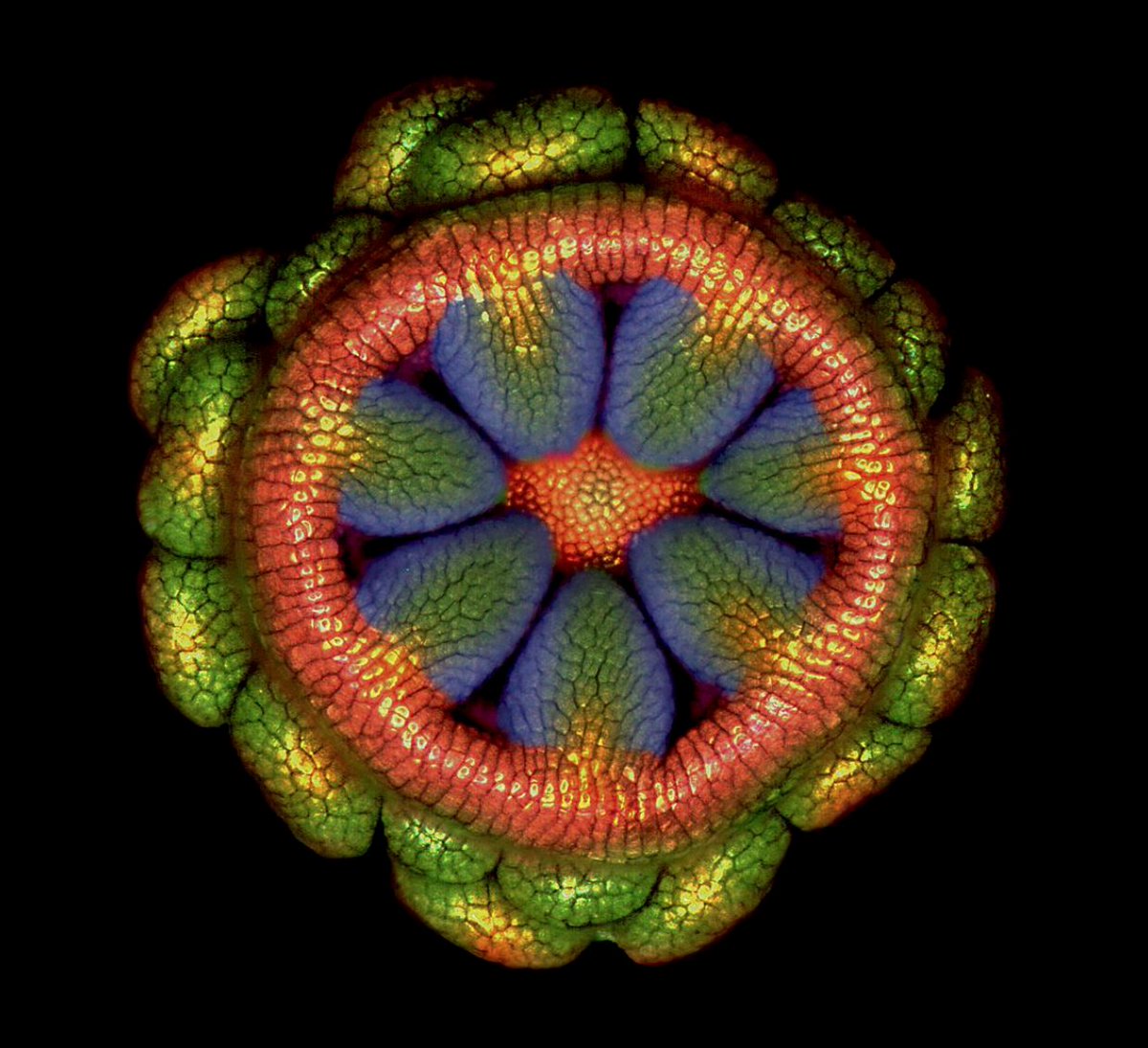
Masoumeh Sahar Khodaverdi's beautiful #microscopy images of #flowers won several awards at the Nikon Small World and @OlympusLifeSci #Bioscapes competitions!

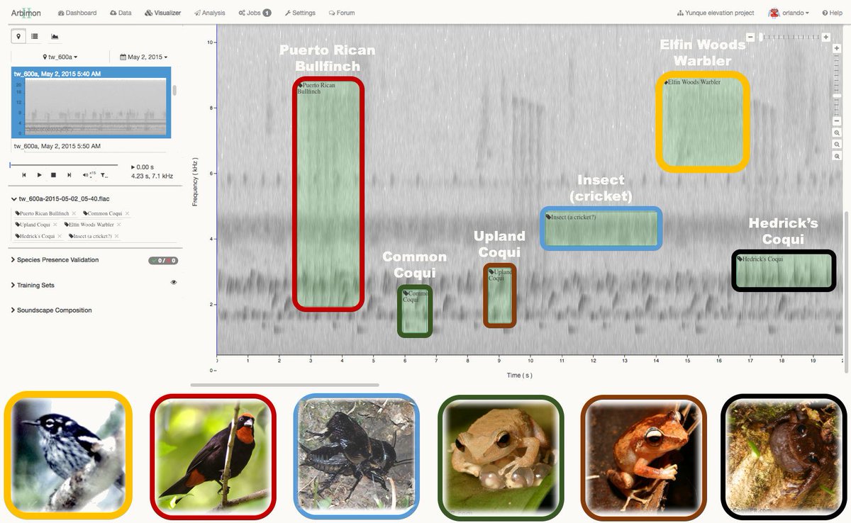
PNASNews Amy McDermott Leah Barclay Soundcamp BiosphereSoundscapes Sonic Environments Locus Sonus Songs Of Adaptation Marc Anderson Bioacoustics and Behavioral Ecology Lab 🔊 Listening to Nature: The Emerging Field of #Bioacoustics —'Researchers are increasingly placing microphones in forests and other ecosystems to monitor birds, insects, frogs, and other animals.'— Cool article on studying natural #soundscapes . bit.ly/2OMDudd Yale Environment 360

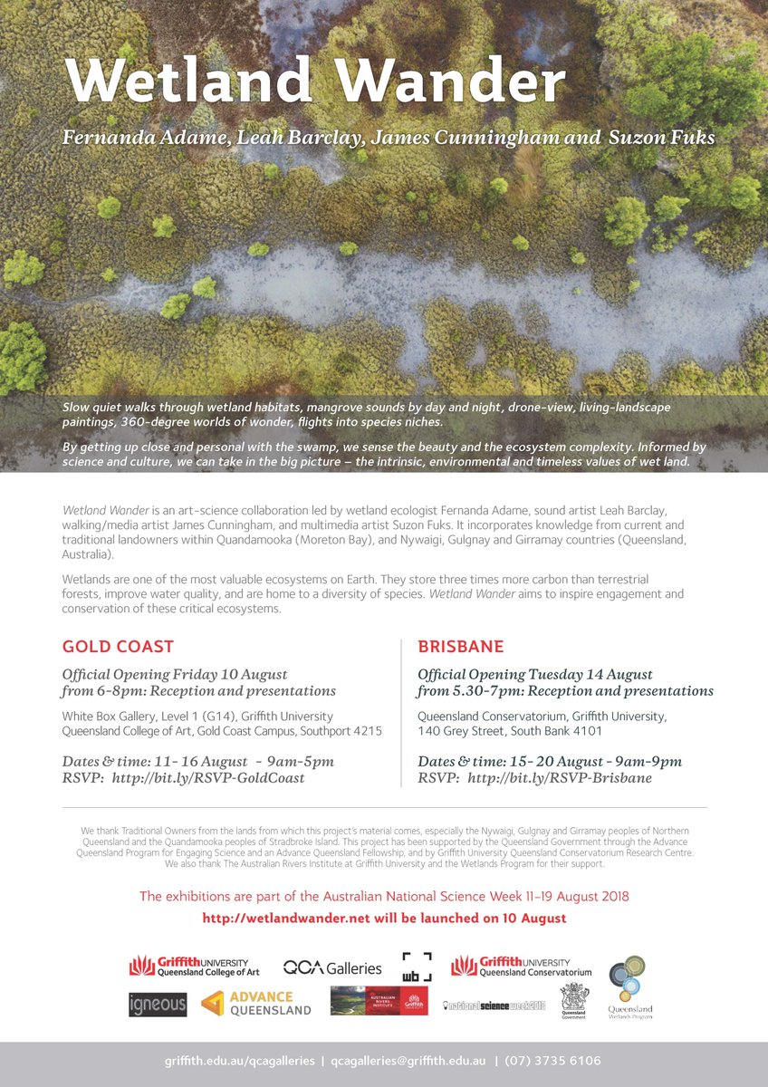
#wetlandwander is an art/science exhibition being launched on the 10th of August!!!
An exciting project which see's Australian Rivers Institute's Fernanda Adame work with suzon, Leah Barclay and BiosphereSoundscapes.
Griffith University @Griffith_ENV @GU_Sciences Griffith HDR Qld Conservatorium.

Biosphere Soundscapes presentation this morning with Leah Barclay at the World Forum for Acoustic Ecology conference in the USA #acousticecology #ecoacoustics

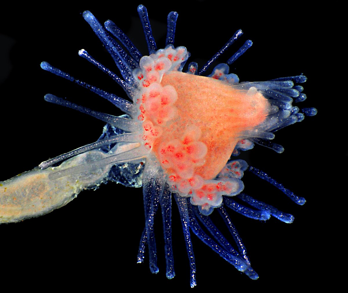
🔬Looking to be dazzled on this #MicroscopyMonday ? Check out this beautiful hydroid collected from a kelp sample
📷: Mike Crutchley/2011 Olympus BioScapes Digital Imaging Competition


![[n]Beyin (@nBeyin) on Twitter photo 2018-08-31 11:33:09 2004 Olympus BioScapes Birincisi: Gözün Mikroskobik Görüntüsü
Donald Pottle tarafından elde edilen bu mikroskobik görüntüde, göz arteriyolleri, esnek elastin duvar (pembe), kırmızı kan hücreleri(kırmızı) ve destekleyici kollajen lifler görünüyor(sarı/yeşil alanlar). (40x obj) 2004 Olympus BioScapes Birincisi: Gözün Mikroskobik Görüntüsü
Donald Pottle tarafından elde edilen bu mikroskobik görüntüde, göz arteriyolleri, esnek elastin duvar (pembe), kırmızı kan hücreleri(kırmızı) ve destekleyici kollajen lifler görünüyor(sarı/yeşil alanlar). (40x obj)](https://pbs.twimg.com/media/Dl7NEG2XoAEqEq_.jpg)
