Semesta Sains(@semestasains) 's Twitter Profile Photo

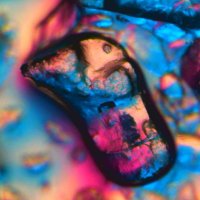
Ada apa di ujung (nyaris di ujung) ballpoint pen.

Teknologi sains kini sudah tidak lagi terbatas pada Electron Microscope, tapi sudah menggunakan Atomic force microscopy (Mikroskop atom).

Video di bawah menggunakan Electron Microscope.

account_circle
RMS(@RoyalMicroSoc) 's Twitter Profile Photo

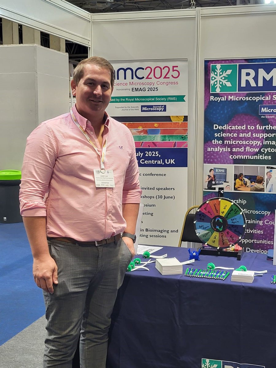
Great to be at the Advanced Materials Show 2024 in Birmingham! Advanced Materials Show

Come and visit our stand to find out more about the world's oldest Society dedicated to !

Great to be at the Advanced Materials Show 2024 in Birmingham! @MaterialsShow #AMS24 #ACS24

Come and visit our stand to find out more about the world's oldest Society dedicated to #microscopy!
account_circle
THE TAO OF NOW(@InTheNoosphere) 's Twitter Profile Photo

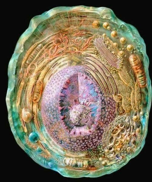
This photo is the most detailed model of a human cell to date, obtained using X-ray, NMR, and cryo-electron microscopy data

This photo is the most detailed model of a human cell to date, obtained using X-ray, NMR, and cryo-electron microscopy data
account_circle
Physics In History(@PhysInHistory) 's Twitter Profile Photo

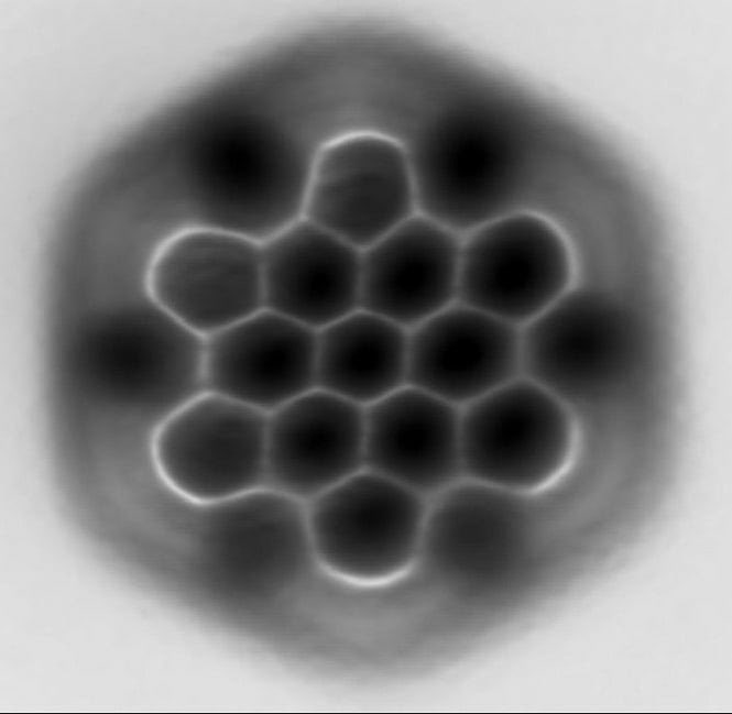
This is an actual picture of a molecule taken by IBM created by using a technique called Atomic Force Microscopy (AFM).

(Credit: IBM)

This is an actual picture of a molecule taken by IBM created by using a technique called Atomic Force Microscopy (AFM).

(Credit: IBM)
account_circle
PicoQuant(@PicoQuant) 's Twitter Profile Photo

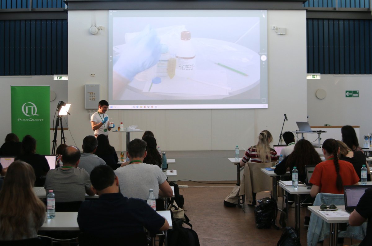
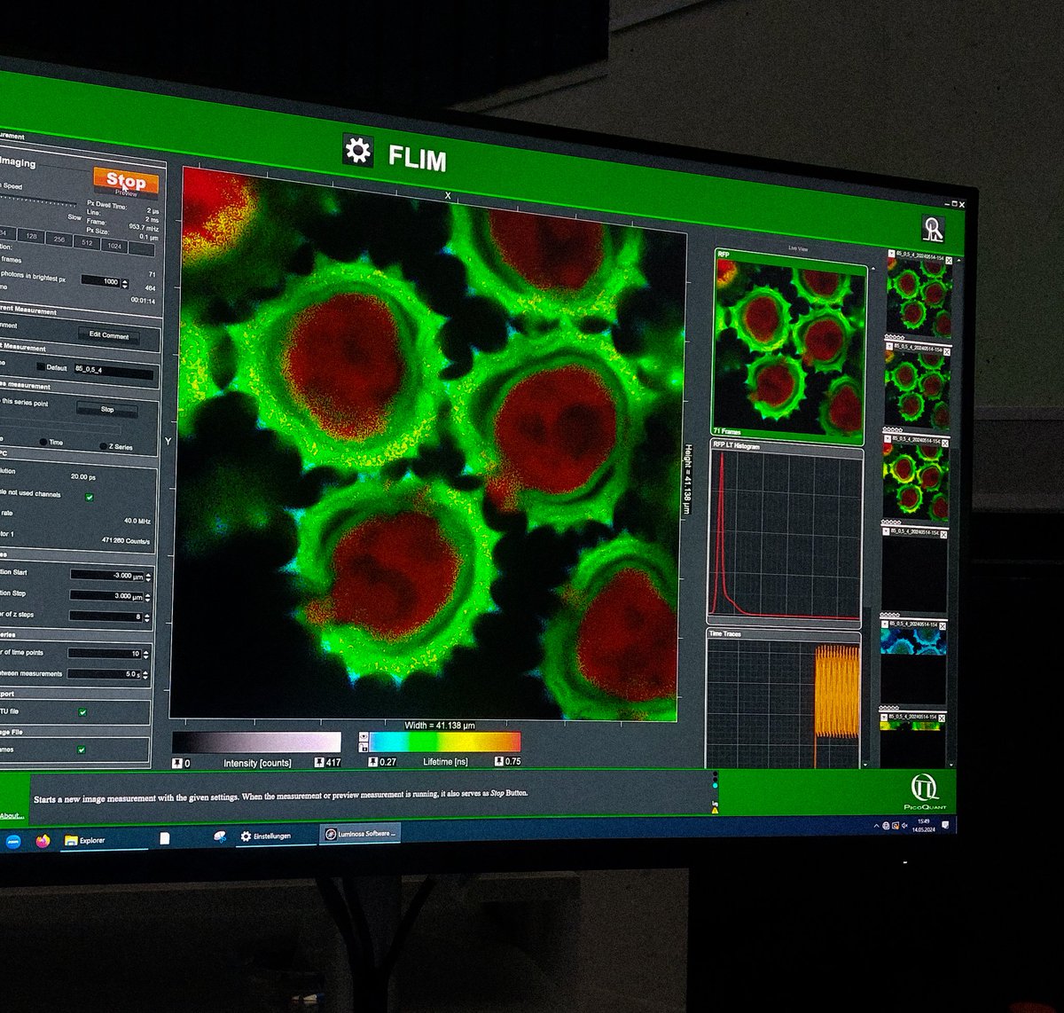
40 highly motivated international scientists have come together in Berlin to participate in our 16th international time-resolved Microscopy Course. This will be a very exciting event!

Our course provides an in-depth introduction to the world of time-resolved fluorescence…

40 highly  motivated international scientists have come together in Berlin to participate in our 16th international time-resolved Microscopy Course.  This will be a very exciting event!  

Our course provides an  in-depth introduction to the world of time-resolved fluorescence…
account_circle
Danielle Beckman(@DaniBeckman) 's Twitter Profile Photo

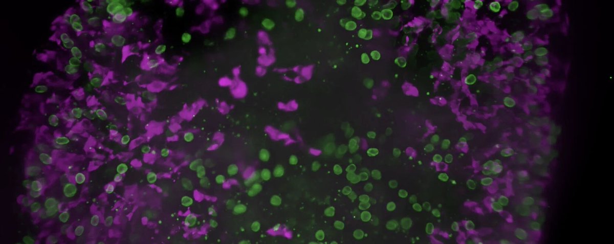
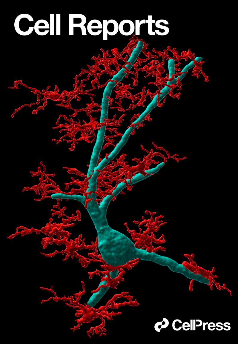
In 2022, we published our first study about developing a new monkey model of . The image below is a 3D reconstruction of a microscopy image showing microglia cells (🔴iba1) attacking a neuron (🔵neurofilament) infected with SARS-Cov-2 (not shown). Do you want to see more?…

In 2022, we published our first study about developing a new monkey model of #COVID19. The image below is a 3D reconstruction of a microscopy image showing microglia cells (🔴iba1) attacking a neuron (🔵neurofilament) infected with SARS-Cov-2 (not shown). Do you want to see more?…
account_circle
Eric Betzig(@Eric_Betzig) 's Twitter Profile Photo


A neutrophilic cell migrating through a 3D collagen mesh at 1.3 sec intervals for 5.5 min, as seen by lattice light sheet microscopy. doi.org/10.1126/scienc…

account_circle
Microscopy Society(@MicroscopySoc) 's Twitter Profile Photo

Come for the Science and Stay for the Wineries! 🍇🍷 Many vineyards are within 1 hour of Cleveland! Register Now: ow.ly/Kw0S50QXVIJ

Learn about the wineries: ow.ly/76q150R8oLm

Come for the Science and Stay for the Wineries! 🍇🍷 Many vineyards are within 1 hour of Cleveland! Register Now: ow.ly/Kw0S50QXVIJ #MM2024 #Microscopy #DiscoverCleveland 

Learn about the wineries: ow.ly/76q150R8oLm
account_circle
Dylan Burnette(@MAG2ART) 's Twitter Profile Photo

A cell undergoing cell division videoed through a microscope. Chromosomes are shown in pink. Differential Interference Contrast (DIC) Microscopy is shown in cyan.

account_circle
Sara Corbezzolo(@saracorbb) 's Twitter Profile Photo

Juanma García-Arcos adding some fun ✨ to this first day of the 16th International Course on 'Time-resolved Microscopy and
Correlation Spectroscopy' 🔬
organized by PicoQuant in Berlin.

@juanmagararc adding some fun ✨ to this first day of the 16th International Course on 'Time-resolved Microscopy and
Correlation Spectroscopy' 🔬
organized by @PicoQuant in Berlin.
account_circle
Killer.T, RMT(@Katusok_RMT2024) 's Twitter Profile Photo

‼️ MTLE Reviewees ‼️

Clinical Microscopy Assessment: forms.gle/hHsvRg4xsZQ2UK…

Please RETWEET to reach MTLE reviewees. Thank you 😊🫶🏻

‼️ MTLE Reviewees ‼️

Clinical Microscopy Assessment: forms.gle/hHsvRg4xsZQ2UK…

Please RETWEET to reach MTLE reviewees. Thank you 😊🫶🏻
account_circle
Physics In History(@PhysInHistory) 's Twitter Profile Photo

This is the most detailed model of a human cell to date, obtained using x-ray, NMR and cryoelectron microscopy datasets.

‘Cellular landscape cross-section through a eukaryotic cell.’ - by Evan Ingersoll and Gael McGill.

This is the most detailed model of a human cell to date, obtained using x-ray, NMR and cryoelectron microscopy datasets. 

‘Cellular landscape cross-section through a eukaryotic cell.’ - by Evan Ingersoll and Gael McGill.
account_circle