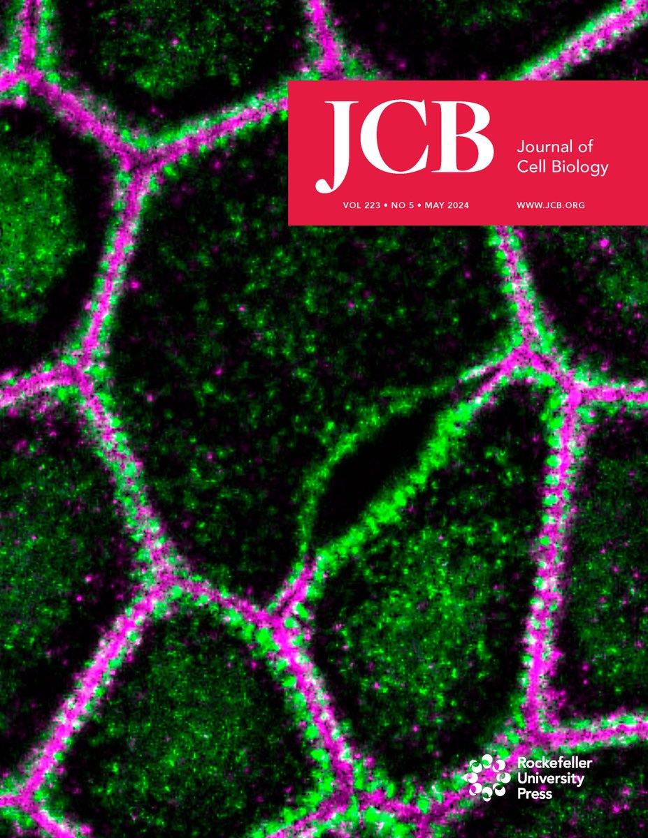
Our May Issue is out! ➡️ hubs.la/Q02vLlxR0
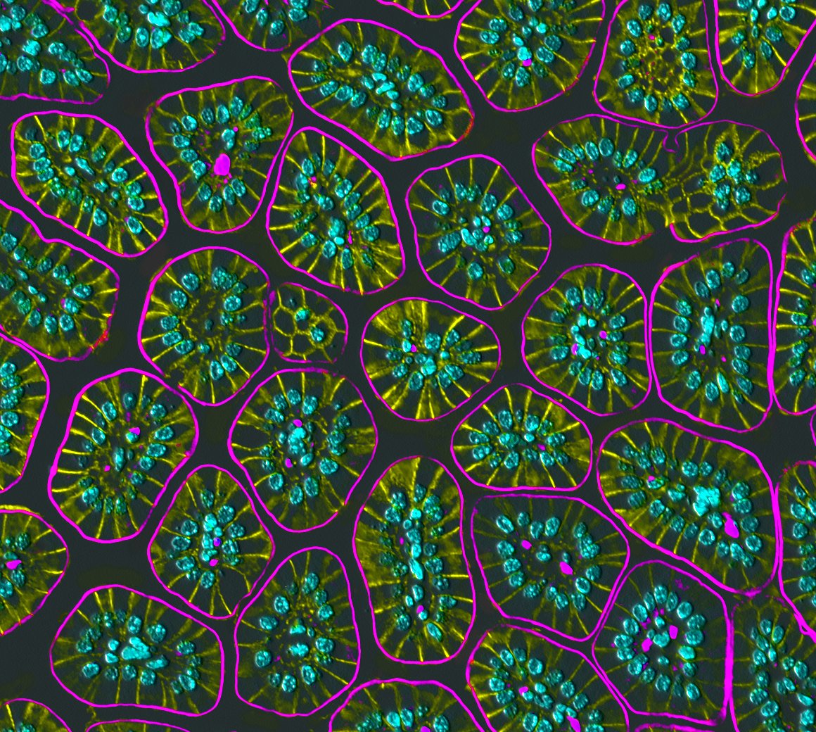
The cover shows STED microscopy image of myosin IIA (green) and ZO-1 (magenta) of claudin/JAM-A KO MDCK II cells. From Thanh Phuong NGUYEN (tann), Tetsuhisa Otani et al. (hubs.la/Q02vLjf10)

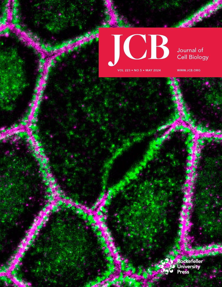
.JCellBiol's May Issue is out! ➡️ hubs.la/Q02vLmCj0
The cover shows STED microscopy image of myosin IIA (green) and ZO-1 (magenta) of claudin/JAM-A KO MDCK II cells. From Thanh Phuong NGUYEN (tann), Tetsuhisa Otani et al. (hubs.la/Q02vLhrT0)
I'm not ready for a new work week but I do enjoy all of the #MicroscopyMonday twitter images.
#bioart #sciart #pathart #histoart #microscopy #intestine
Good Morning/Afternoon Everyone!
☀️🌅✨
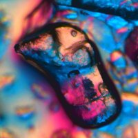
Dive into the micro salt realms!🧂🔬
Over 600 #NFTArtworks 📸🎨
#NFTPhotography #Microscopy
Visit now!
opensea.io/assets/ethereu…

💎”tsunami on the moon”💎
1/600+ #NFTPhotography #NFTArtworks #Microscopy
Dive into the beautiful and unique world of the micro salt realms⭐️
opensea.io/assets/ethereu…

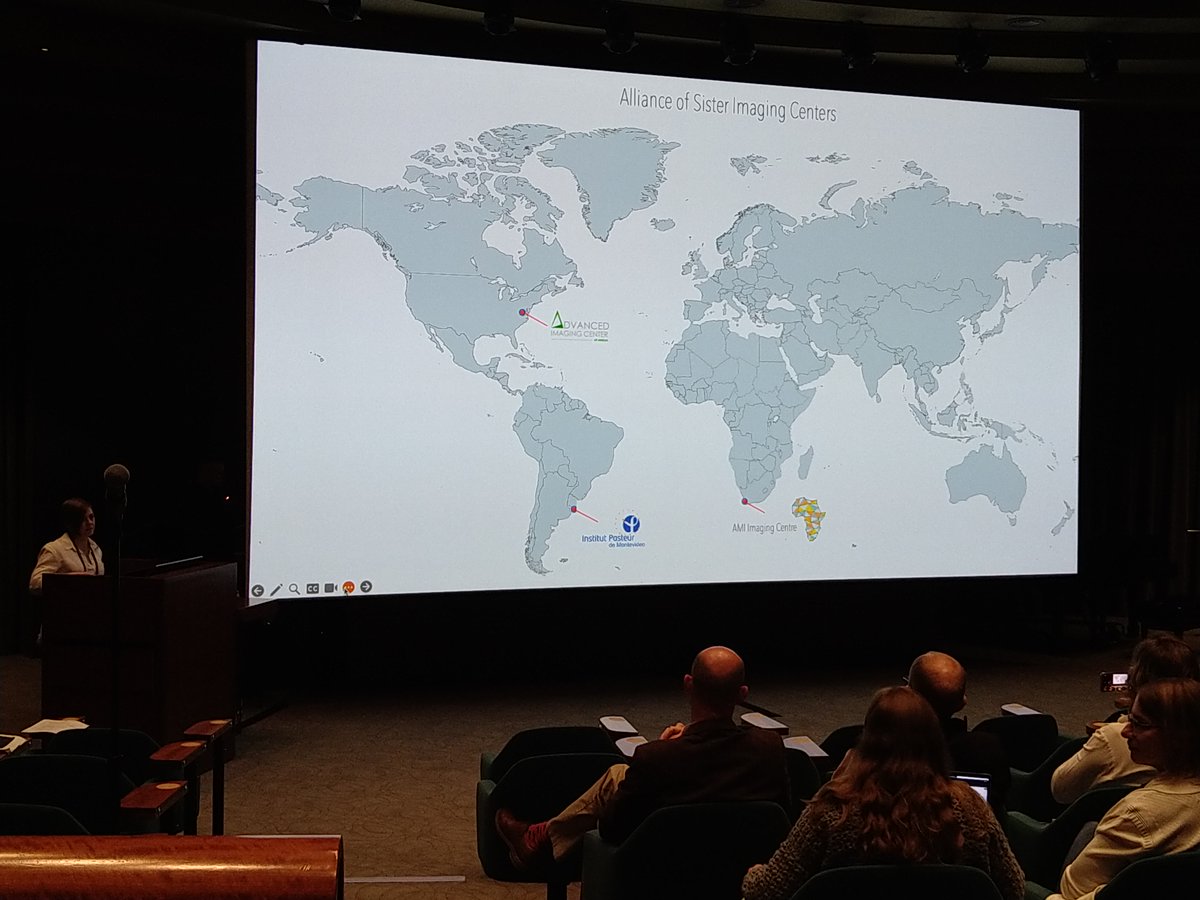
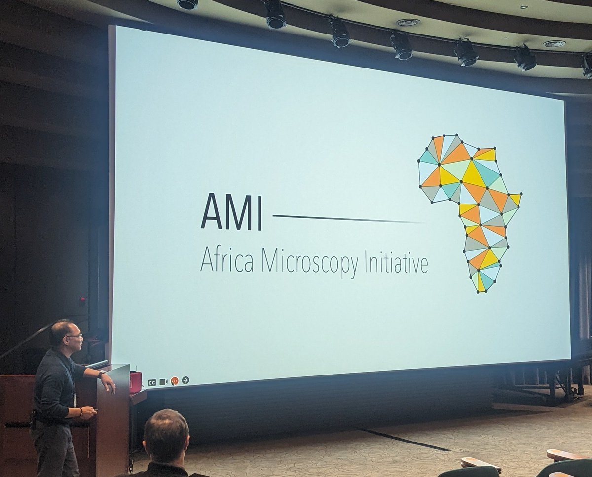
Fantastic welcome from the organisers of the Janelia Conferences on disseminating microscopy technology to underserved communities. We're excited to learn more from this diverse community and find out how we can contribute!
#SpreadingMicroscopy

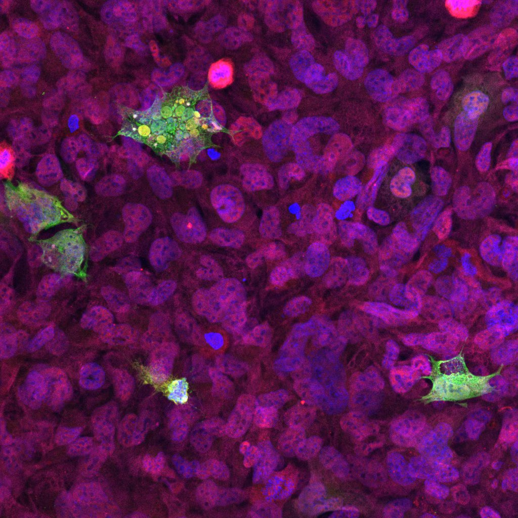
Happy Fluorescence Friday! 🧪
GFP stained human microglia! 🤓
#fluorescencefriday
#microscopy
#neuroscience

Can't believe it's been 5 years since I started my #Python tutorial channel! From learning new topics to almost hitting 100K subscribers, it's been an incredible journey. Massive thanks to all my subscribers!
#Microscopy #DeepLearning #Bioimageanalysis

#FluorescenceFriday #cellbiology #bioart Mitochondrial dynamics in a cultured pig kidney cell (LLC-PK1) at 3 volumes/min for 97 min by two-photon Bessel beam plane illumination microscopy. doi.org/10.1038/nmeth.…

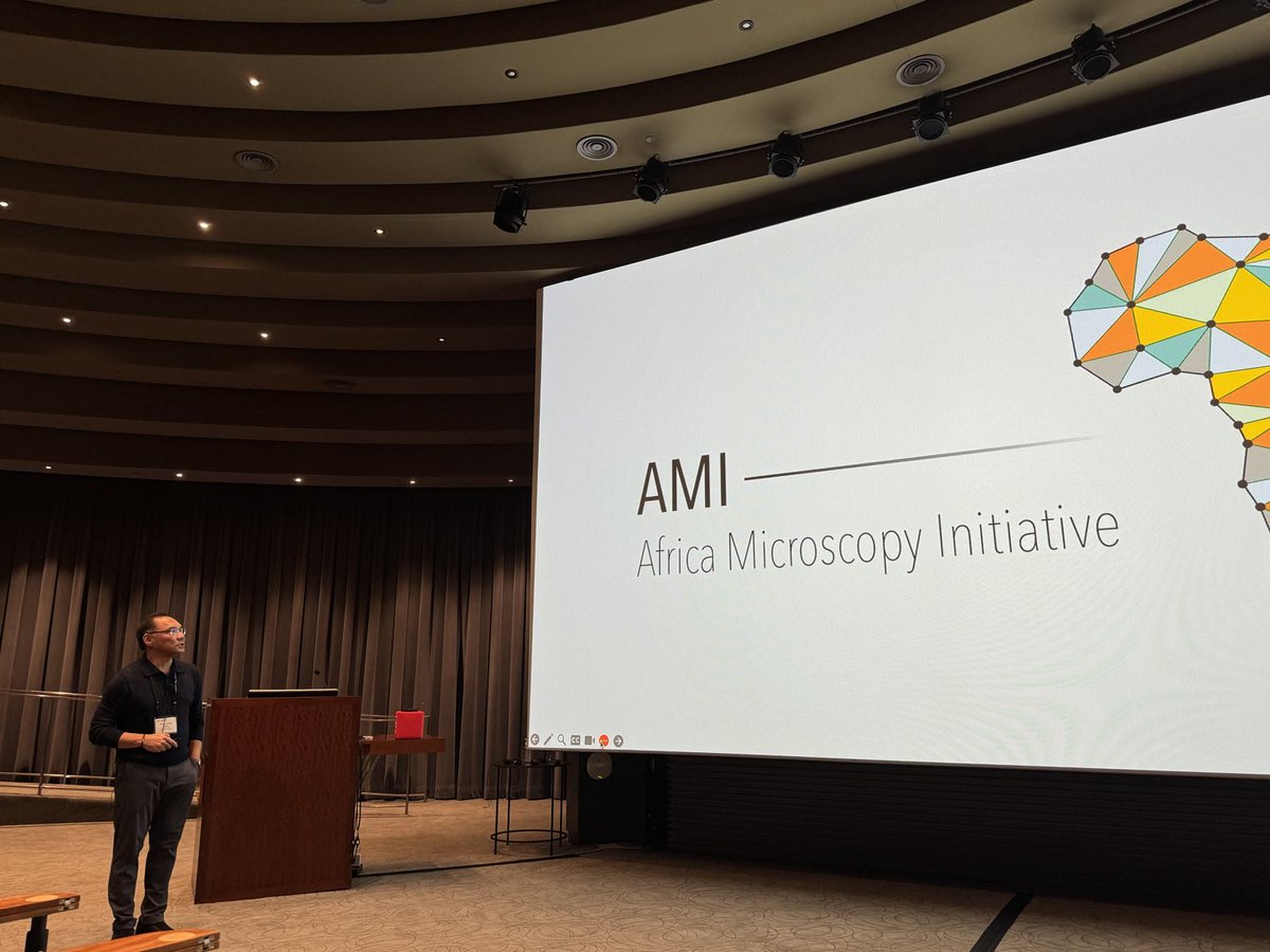
Leong kicking off the #spreadingmicroscopy meeting! Africa Microscopy Initiative HHMI | Janelia AIC at Janelia

Enter the #microscopic terrains of salt🔬
⭐️”A half”⭐️-make an offer now!
🟢600+ #Microscopy #NFT Artworks
#NFT Photography #NFT Artist #NFT opensea.io/assets/ethereu…

Late GM/GA Everyone!✨☀️
Embark on a journey ✨🧂🔬
into the Micro-Salt realms!
Over 600 #NFTArtworks
#NFTPhotography #Microscopy #OpenseaNFT #NFTCommunity #NFTCollection
opensea.io/assets/ethereu…

Some fun non- #lightsheet #Microscopy 🔬: A polychaete imaged at Marine Biological Laboratory (MBL)


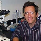
Teng-Leong Chew 周呈隆 kicking things off for Janelia's Microscopy Dissemination conference #SpreadingMicroscopy ! @HHMI

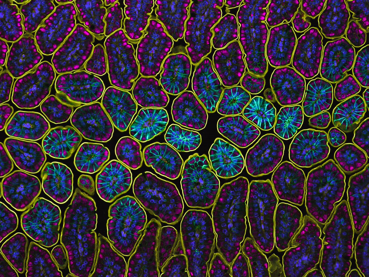
Claudin 4 (teal) is such a wild claudin...
#FluorescenceFriday #FluorescentFriday #pathart #bioart #sciart #intestine #histoart #microscopy

Happy Wednesday Afternoon🌅⭐️✨🧂🔬 #Microscopy #NFTPhotography
Enter the micro salt realms!👇
opensea.io/assets/ethereu…
